FLOW CHART IS A GUIDELINE ONLY
Dry Cough?
Shortness of breath?
Fever >37.9C?
Pneumonia suspected or already diagnosed (imaging positive)?
…or other clinical suspicion of Covid-19
Aims:
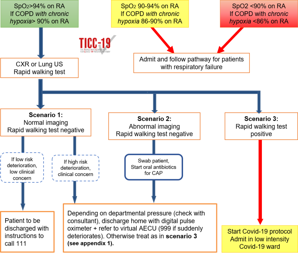
- Identify suspected Covid-19 patients who do not require hospital admission.
- Start intensive therapy pathways for those with respiratory insufficiency.
Appendix 1
Rapid Walking Test
Take baseline oxygen saturations. Encourage the patient to walk at the highest possible speed for 30 meters on a linear path. Sit the patient down and re-measure oxygen saturations. The test is considered positive if the absolute oxygen saturation has fallen by more than 5%.
A note on patients with COPD.
Many patients with COPD do not have sats of 88-92% but do in fact have normal sats, so do not assume that the patient with low sats who has COPD represents a patient with ‘normal’ sats for them. They may be severely unwell. If in doubt, do an ABG or admit.
Lung Ultrasound
Lung ultrasound should only be used in place of a chest x ray by clinicians confident in their lung ultrasound ability. 12 point lung ultrasound. If A lines (horizontal lines) in all zones then lung ultrasound ‘normal’ (ie scenario 1). If any abnormality (B lines, consolidation) then considered ‘abnormal’ (ie scenario 2).
Adaptation of pathway for inpatient use.
Patients assessed as inpatients on medical wards may be able to go through the same triage pathway to be discharged (ie walking test negative but imaging changes to go home with a sats probe and monitored through ambulatory).